Implants & abutments
Use matched photogrammetry scanbodies to record the relative implant or MUA positions.
PhotogrammetryAtomica photogrammetry
Record the relative positions of implants or multi-unit abutments. Connect the capture to your full-arch restorative workflow.
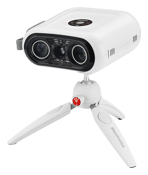 A dedicated view of the implant positions.
Built around full-arch work
Photogrammetry captures the implant coordinates. Combine them with surface scans and bite records to give your restorative team the information it needs.
Use matched photogrammetry scanbodies to record the relative implant or MUA positions.
PhotogrammetryCapture the surface anatomy with your intraoral scanner and add other case records as needed.
Intraoral scanningInclude bite records and relevant prosthesis scans to support alignment and restorative design.
Case recordsFrom capture to restoration
Confirm implant or MUA compatibility, fit the matched scanbodies and capture the required measurement points.
Export the position data and combine it with the intraoral scans, bite records and other required case information.
Send the case to a compatible CAD workflow and matching library for alignment, restorative design and subsequent verification.
Across the restorative journey
Provisional workflow
Combine implant position data with the relevant scans and bite information to support the design of a full-arch provisional restoration.
Definitive workflow
Provide updated tissue records and reference prosthesis scans, as needed, alongside implant position data for the definitive restoration.
System specifications
A dedicated dual-camera system for measuring implant scanbody positions. Confirm the scanbody and CAD library combination for your implant system before ordering.
Manufacturer measurement specifications. Clinical results depend on the complete capture, alignment, design and verification workflow.
A few useful answers
Yes. Photogrammetry supplies implant position data. An intraoral scanner supplies tissue and opposing-arch surfaces, alongside the bite and other records required for the restorative case.
Compatibility depends on the implant or multi-unit abutment, matched photogrammetry scanbodies and the CAD library. Share your system and restorative workflow with our team so we can confirm the correct combination.
The system supports STL export for compatible CAD workflows, including workflows using exocad. Your lab should confirm the library and record requirements before the first case.
See the current offer for the camera, accessories, scanbody options, onboarding and warranty. Our team can help configure the package for your implant systems.
See it in your workflow
Walk through the capture process with our team and confirm compatibility with your implant systems and lab.
Connected by your workflow